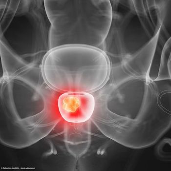
FDA accepts application for 18F-rhPSMA-7.3 for prostate cancer
The application is supported by findings from a phase 1 trial and two phase 3 trials, SPOTLIGHT and LIGHTHOUSE.
The FDA has accepted a new drug application (NDA) for the radiohybrid PSMA-PET imaging agent 18F-rhPSMA-7.3for diagnostic imaging of prostate cancer, according to Blue Earth Diagnostic, the developer of the agent.1
The NDA is supported by findings from a phase 1 trial and two phase 3 trials, SPOTLIGHT and LIGHTHOUSE. The SPOTLIGHT data were shared at medical conferences earlier this year; however, the findings from LIGHTHOUSE have not yet been made available.
“Prostate cancer is a leading cause of male cancer-related death worldwide, and accurate localization and staging of the disease is critical in establishing optimal medical management strategies,” Eugene Teoh, MBBS, MRCP, FRCR, DPhil, chief medical officer of Blue Earth Diagnostics, stated in a press release. “We believe that the performance of 18F-rhPSMA-7.3, its high PSMA binding affinity and potential for low bladder activity will make it a valuable diagnostic tool that is radiolabeled with 18F for high image quality and readily available patient access.”
SPOTLIGHT Data
The latest data from the phase 3 SPOTLIGHT trial presented earlier this year at the 2022 AUA Annual Meeting showed that compared with baseline conventional imaging, PET imaging with 18F-rhPSMA-7.3 frequently led to post-scan disease upstaging in men with prostate cancer recurrence.2
Among 250 of 366 men in the efficacy analysis population who had negative baseline conventional imaging, 18F-rhPSMA-7.3 showed a correct detection rate between 45% and 47%, which is the percentage of patients scanned with at least one true positive PET finding compared with the Standard of Truth of histopathology or confirmatory conventional imaging. Notably, upstaging results were found to vary based on prior treatment and anatomical region.
Initial data from SPOTLIGHT (NCT04186845) previously presented at the 2022 Genitourinary Cancers Symposium showed that the overall detection rate in patients who had an evaluable 18F-rhPSMA-7.3 scan was 83% by majority read.3
SPOTLIGHT enrolled male patients who were at least 18 years of age and received prior curative intent treatment for localized prostate cancer. Patients were required to have elevated PSA that could indicate biochemical recurrence and be eligible for salvage therapy.
18F-rhPSMA-7.3 was administered at a dose of 296 MBq/8 mCi, and PET/CT scans were given between 50 and 70 minutes following injection. Scans were then evaluated by 3 blinded central readers. The Standard of Truth was histopathology or confirmatory conventional imaging. Biopsies took place with 60 days following PET scans, and confirmatory imaging was conducted within 90 days.
Of the 420 patients who consented for the trial, 391 received 18F-rhPSMA-7.3, of which 389 underwent an evaluable PET/CT scan. Furthermore, 366 patients had sufficient data to determine Standard of Truth; these patients comprised the efficacy-evaluable population. Specifically, 69 patients had Standard of Truth determined by histopathology compared with imaging in 297 patients.
In this population, the mean age was 68.4 years (range, 43-85). The median PSA was 1.27 ng/mL (range, 0.03-134.6), and most patients had a Gleason grade group of 3 (30%).
Additional data showed the 18F-rhPSMA-7.3 detection rate was 88% compared with a correct detection rate of 57%.
In patients with negative baseline conventional imaging in the prostate bed region, between 3.5% and 8.0% of patients post prostatectomy showed true positive detections compared with between 39% and 41% of patients post radiation therapy.
In patients with negative baseline conventional imaging in the pelvic lymph node region, between 18% and 21% of patients post prostatectomy showed true positive detections vs 6.5% of patients post radiation. In patients with negative baseline conventional imaging in the extra-pelvic region, between 21% and 26% of patients post prostatectomy showed true positive detections vs between 20% and 30% of patients post radiation.
"This event marks a significant milestone in advancing our robust prostate cancer portfolio, and we are very pleased that the FDA has accepted our NDA submission for the use of 18F-rhPSMA-7.3 PET imaging in prostate cancer patients," David E. Gauden, DPhil, chief executive officer of Blue Earth Diagnostics, stated in this press release
"We look forward to working with the Agency throughout the review process, with the goal of having an approved product that is widely available and accessible across the United States. Subject to FDA approval, we believe that 18F-rhPSMA-7.3 PET imaging may be clinically useful in the management of men affected by prostate cancer across the care continuum. All of us at Blue Earth want to express our sincere gratitude to the many patients, physicians, clinical trial sites and collaborators who have worked closely with us to progress 18F-rhPSMA-7.3, and to having successfully completed our phase 3 clinical trials despite all the challenges presented by the COVID-19 pandemic," added Gauden.
More on SPOTLIGHT:
References
1. Blue Earth Diagnostics Announces FDA Acceptance of New Drug Application for 18F-rhPSMA-7.3, a Radiohybrid Prostate-Specific Membrane Antigen-Targeted PET Imaging Agent for Prostate Cancer. Posted online September 28, 2022. Accessed September 29, 2022. https://bwnews.pr/3y4rbRf
2. Fleming MT. Impact of 18F-rhPSMA-7.3 PET on upstaging of patients with prostate cancer recurrence: results from the prospective, phase 3, multicenter, SPOTLIGHT study. Presented at: 2022 AUA Annual Meeting; May 13-16, 2022; New Orleans, LA. Abstract PLLBA-02.
3. Schuster DM. Detection rate of 18F-rhPSMA-7.3 PET in patients with suspected prostate cancer recurrence: results from a phase 3, prospective, multicenter study (SPOTLIGHT). J Clin Oncol. 2022(suppl 6):9. doi:10.1200/JCO.2022.40.6_suppl.009
Newsletter
Stay current with the latest urology news and practice-changing insights — sign up now for the essential updates every urologist needs.


