- Vol 50 No 09
- Volume 50
- Issue 09
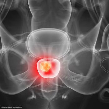
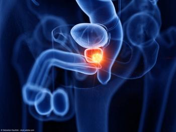
PSMA and MRI for early prostate cancer detection
Experts weigh in on whether the combined technologies could be the future power couple of prostate cancer diagnosis.
Prostate-specific membrane antigen (PSMA) and multiparametric magnetic resonance imaging (MRI) are proving their worth, individually, in the diagnosis and treatment of prostate cancer. But each has its limitations and PSMA use is indicated only for advanced prostate cancer.
Now investigators are asking whether marrying the imaging capabilities of PSMA positron emission tomography (PET) and MRI might optimize use of imaging while limiting the need for invasive biopsies.
“PSMA has an opportunity to potentially delineate areas of the prostate where the MRI can miss cancer. We know there is no perfect imaging modality that has 100% specificity and 100% sensitivity. So to layer in a PET scan like PSMA with MRI has the potential to find additional tumors. The caveat to this is while it is what we believe and hope to happen, until we have definitive clinical trials that show this, we cannot assume that’s the case,” said Peter A. Pinto, MD, an investigator and urologic senior surgeon in the Urologic Oncology Branch of the National Cancer Institute, National Institutes of Health, Bethesda, Maryland.
Rapidly improving imaging for prostate cancer, from new MRI and ultrasound technologies to integrated, multiparametric imaging and image fusion such as PSMA/MRI, will not only affect clinicians’ ability to identify and characterize tumors within the gland but also will play a central role in risk stratification for patients and treatment decision-making, according to Jonathan Coleman, MD, professor and surgeon in the Department of Surgery/Urology, Memorial Sloan Kettering Cancer Center, Weill Cornell Medical Center, New York, New York.
The challenges will be in establishing clinical pathways that allow urologists and others to rely on the accuracy of these tests and understand how to best incorporate them into practice.
“Issues like: Which cases are best suited to these studies? How often should they be performed if using them for follow-up? Are they accurate for confidently identifying or excluding features of extra-prostatic disease?” Coleman said. “These are issues that need to be studied and answered; however, often the cart gets put before the horse—especially when it comes to imaging technologies. Everyone is curious and wants to ‘see’ what is going on with their disease but is not quite sure how to use that information to make clinically relevant, data-driven decisions.”
The technologies, potential benefits of combining them
MRI has a well-established role for men suspected of having prostate cancer, according to Declan G. Murphy, MB, BCh, BAO, FRACS, FRCS (Urol), a consultant urologist and director of genitourinary oncology at the Peter MacCallum Cancer Centre in Melbourne, Australia.
“MRI in men suspected of having prostate cancer is a fantastic tool—fully funded and fully integrated in Australia. Same in the UK. Outside some academic centers, the US has been rather slow to embrace MR in early prostate cancer detection, mostly for financial reasons. But the data are clear and, in every single guideline, MRI is there,” said Murphy, an author of the PRIMARY trial published last year in European Urology.1 “If the MRI looks normal, those men very often don’t need a biopsy and can be reassured. Second, if the MRI is abnormal, then we do a targeted biopsy. This approach is well supported by the data from the PRECISION trial, for example.”2
In December 2020, the FDA approved Gallium 68 PSMA-11 (Ga 68 PSMA-11), the first drug for PET imaging of PSMA-positive lesions in men with prostate cancer.
“The indication for PSMA for advanced cancer has been established,” Pinto said. “PSMA has now been approved to improve the staging of patients with aggressive prostate cancer to evaluate distant disease.”
Although multiparametric MRI (mpMRI) remains the gold-standard imaging modality for diagnosing prostate cancer, PSMA PET/CT was shown to have similar diagnostic accuracy for the identification of prostate cancer foci, said Ida Sonni, MD, project scientist in the Department of Radiology at the David Geffen School of Medicine at the University of California, Los Angeles.
“But mpMRI allows a better soft-tissue evaluation and is superior in the assessment of the local extent of disease, or T staging. PSMA PET and mpMRI provide complementary information and integrating them using PSMA PET/MRI provides significant advantages,” said Sonni, who is lead author of the recent paper “Head-to-Head Comparison of 68Ga-PSMA-11 PET/CT and mpMRI with a Histopathology Gold Standard in the Detection, Intraprostatic Localization, and Determination of Local Extension of Primary Prostate Cancer: Results from a Prospective Single-Center Imaging Trial,” published in the Journal of Nuclear Medicine.3
Using PET/MRI instead of PET/CT should offer a much-improved chance to evaluate the soft tissues of the prostate, both structurally (with MRI) and functionally (with PET), according to Leonard S. Marks, MD, deKernion Endowed Chair in Urology at the David Geffen School of Medicine at the University of California, Los Angeles. Marks was among the first in the US to use MRI/ultrasound fusion for prostate biopsy, while at UCLA in 2009.
One thing that is unlikely to become part of clinical practices is a single device integrating the PET camera with an MR system, according to Murphy.
Promising data with systems integrating the PET camera with MRI rather than CT prompted Murphy to investigate this technology about 8 years ago, when Murphy and his colleague Michael Hofman, MBBS, professor of nuclear medicine at Peter MacCallum Cancer Centre, traveled to Germany to meet with researchers in Munich who had published their experience in using the dual machine.
“The clear, overarching message is that PSMA PET/MR, rather than PET/CT (which is where all the data have been), will never be integrated into clinical practice. It’s too expensive and too inefficient for clinical practice for prostate cancer, Murphy said. “This is especially so in countries like the US that already struggle to do prostate MRI pre-biopsy.”
The solution appears to be in using software to fuse PET and MRI images separately, according to Murphy.
“We routinely fuse PET and MRI images. That’s a better workflow, a better-quality MR … and is a more useful clinical tool. It’s more pragmatic. You can do it in your clinical practice,” Murphy said.
Total-body PET/CT is the most exciting evolution of PET technology today, not PET/MRI, which will never be practical for prostate cancer, according to Murphy.
Evidence needed
Pinto, who was among the investigators to usher in the era of prostate MRI as an established technique for prostate cancer detection, remembers the skepticism about MRI’s value.
“In the early years of prostate MRI, people were skeptical that such an expensive imaging modality had any role in prostate cancer detection or early-stage treatment. I think this is very akin to PSMA today,” Pinto said.
Just like prostate MRI did, PSMA will need clinical trials from multiple centers published in highly respected peer reviewed journals to support its utility in the diagnostic algorithm to detect prostate cancer. Replacing the current standard of care is an arduous task since prostate cancer is a slow-growing cancer—one that has both indolent and aggressive types depending on the cancer grade. Therefore, screening and detection trials require many patients with appropriate follow-up, according to Pinto.
“Long term, the hope is such clinical trials will demonstrate that this new method to detect prostate cancer with PSMA/MRI is actually decreasing the mortality from prostate cancer,” Pinto said.
Although PSMA/MRI might not have the level of evidence to be the new standard of care as recommended by guidelines and professional societies anytime soon, it could become a “community standard” if smaller, single-institution studies have positive results and physicians start to incorporate its use in their practice, according to Pinto.
“PSA is still used to screen for prostate cancer and the elevation of PSA creates clinical suspicion often worthy of further investigation. Both physicians and patients often seek out a prostate MRI as the next step. If the prostate MRI finds an abnormality worrisome for a cancer, a prostate biopsy is performed directed to that lesion. In addition, other regions of the prostate where the MRI does not see anything concerning for cancer are biopsied since MRI can miss cancer,” Pinto said. “But what if that biopsy fails to demonstrate cancer yet the urologist is still concerned that there may have been a tumor missed? I can easily see a time when researchers determine if a PSMA/MRI could help detect missed cancers in those areas.”
Payers, according to Marks, won’t like the added expense, initially.
“But when it’s shown to improve accuracy of diagnosis, they will fall into line, just as they have with MRI,” Marks said.
PSMA/MRI with focal treatment?
Decades of research on different types of focal therapy in prostate cancer have revealed a failure or recurrence rate generally between 20% and 40%, according to Pinto.
“Research here to improve focal therapy is focused on the belief that in-field cancer recurrence is not due to the type of ablative modality used; rather, it has to do with being able to see the tumor and its borders well,” Pinto said. “PSMA/MRI may help us to better select patients for focal therapy and help guide or follow up patients in that capacity.”
Early studies
Initial studies that combine PSMA with MRI for prostate cancer diagnosis are promising, according to Pinto.
“But in medicine, often there are early, initial studies that are promising, and only when we look at multiple larger trials do we find out our early belief does not hold true,” Pinto said. “I am cautiously optimistic, though, that as we continue with large-scale studies, we will find PSMA/MRI is a benefit to our patients.”
In the paper, “Imaging Prostate Cancer: Clinical Utility of Prostate-Specific Membrane Antigen,” published in the Journal of Urology, Marks and coauthors write that PSMA PET/CT scanning may help localize cancer within the prostate, citing several studies including the large prospective PRIMARY trial, which compared MRI and PSMA scanning to biopsy results.4
“Emmett and colleagues found the 2 imaging modalities to provide additive information. False negative rate for clinically significant [prostate cancer] was 17% with MRI and 10% with PSMA; but only 5/291 men were falsely negative by both, suggesting the possibility of deferring biopsy in such cases,” the authors wrote. “The value of pre-biopsy imaging, as described in this important paper, deserves further study.”4
In the future, fusing the 2 could create a less invasive diagnostic pathway for prostate cancer, according to Coleman.
“Potentially, and likely along with some other corroborating biomarker from serum or urine,” he said. “[W]e all would like to see a less invasive means for screening men for prostate cancer, though I suspect biopsy will still have a role for firmly establishing the presence of cancer. Most likely the use of biopsy will be much more selective and with only a few samples taken.”
References
1. Emmett L, Buteau J, Papa N, et al. The additive diagnostic value of prostate-specific membrane antigen positron emission tomography computed tomography to multiparametric magnetic resonance imaging triage in the diagnosis of prostate cancer (PRIMARY): a prospective multicentre study. Eur Urol. 2021;80(6):682-689. doi:10.1016/j.eururo.2021.08.002
2. Kasivisvanathan V, Rannikko AS, Borghi M, et al. MRI-targeted or standard biopsy for prostate-cancer diagnosis. N Engl J Med. 2018;378(19):1767-1777. doi:10.1056/NEJMoa1801993
3. Sonni I, Felker ER, Lenis AT, et al. Head-to-head comparison of 68Ga-PSMA-11 PET/CT and mpMRI with a histopathology gold standard in the detection, intraprostatic localization, and determination of local extension of primary prostate cancer: results from a prospective single-center imaging trial. J Nucl Med. 2022;63(6):847-854. doi:10.2967/jnumed.121.26239
4. Kuppermann D, Calais J, Marks LS. Imaging prostate cancer: clinical utility of prostate-specific membrane antigen. J Urol. 2022;207(4):769-778. doi:10.1097/JU.0000000000002457
Articles in this issue
over 3 years ago
Is seminal vesiculectomy included within the prostatectomy code?over 3 years ago
Call it by any name: Bladder that does not workover 3 years ago
How to use life insurance to pay estate taxesover 3 years ago
Do 5-ARIs protect against death from prostate cancer?almost 4 years ago
Radiation therapy for prostate cancer continues to evolveNewsletter
Stay current with the latest urology news and practice-changing insights — sign up now for the essential updates every urologist needs.


